Immunohistochemical Investigation of Cyclooxygenase-2 Expression in Rabbit Uterine Adenocarcinoma and the Potential Use of COX-2 Inhibitors in Cancer Therapy
Authors: Chiara Vaccaro, Sara Meoli, Giuseppe Sarli, et al.
Affiliations: Department of Veterinary Medical Sciences, University of Bologna, Italy.
Investigate COX-2 expression across rabbit uterine adenocarcinoma ($n=30$), endometrial hyperplasia ($n=7$), and normal endometrium ($n=10$) to assess diagnostic value & therapeutic feasibility.
100% of adenocarcinomas expressed COX-2 (mean IHS $8.27 \pm 2.86$), significantly higher than normal endometrium ($0.40 \pm 0.70$) and hyperplasia ($1.71 \pm 1.11$) with $p < 0.001$.
Post-surgical Meloxicam treatment (0.5 mg/kg BID $\times$ 7 days) achieved 100% 18-month metastasis-free survival versus 5.4 months mean survival in surgery-alone controls.
Interactive Visualizations & Statistical Summaries
Data charts illustrating tumor subtype distributions, FIGO grades, COX-2 immunoreactivity scores, and survival rates.
Subtypes & Invasion Depth n = 30 cases
Breakdown of growth pattern, invasion depth, and mitotic count.
FIGO Histological Grade Distribution FIGO Proportions
Proportion of Grade 1, Grade 2, and Grade 3 tumors.
COX-2 IHS Staining Intensity Adenocarcinoma
Weak (1-4), Moderate (6-8), and Strong (9-12) IHS distribution.
COX-2 Immunoreactive Score (IHS) Across Tissue Classifications p < 0.001
Comparison of mean IHS values between normal endometrium, hyperplasia, and adenocarcinoma histological grades.
Survival Analysis: Meloxicam vs Surgery Alone 18-Mo Follow-Up
Comparing post-surgical Meloxicam treatment vs surgery alone mean survival.
Figure 1 Histological Subtypes of Rabbit Uterine Adenocarcinoma
H&E staining illustrating major histopathological growth patterns in domestic rabbits (Click image to zoom).
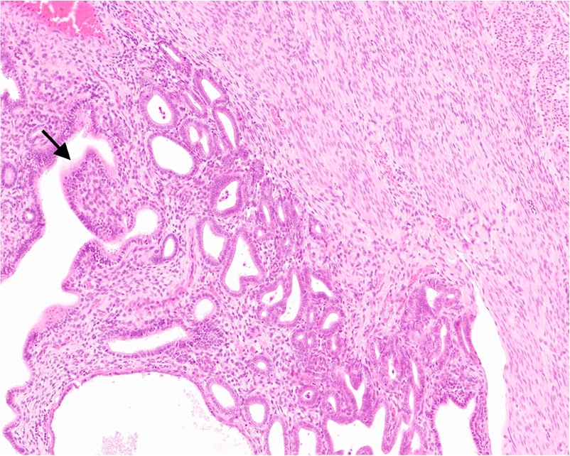 1a
Papillary Subtype
H&E stain showing well-differentiated columnar neoplastic epithelium arranged in complex, branching arborizing papillary fronds supported by delicate vascularized stromal cores.
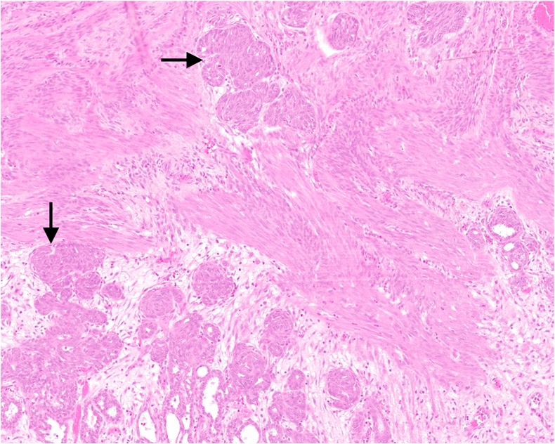 1b
Tubular / Solid Subtype
H&E stain illustrating packed irregular tubular structures transitioning into solid sheets and compact nests of atypical epithelial cells with moderate-to-severe nuclear pleomorphism.
"Uterine adenocarcinomas ($n = 30$) were classified histopathologically according to dominant growth patterns into two distinct subtypes: Papillary (Figure 1a) and Tubular/Solid (Figure 1b). The papillary subtype was identified in 53.3% of cases (16/30), characterized by delicate arborescent papillary projections lined by single or pseudostratified columnar epithelial cells with uniform nuclei. The tubular/solid subtype constituted 46.7% of cases (14/30), displaying crowded, back-to-back gland-like structures or solid sheets devoid of lumen formation."
Myometrial Invasion Data: Superficial myometrial invasion (<50% of wall thickness) was detected in 60% (18/30) of cases, whereas deep myometrial invasion ($\ge 50\%$ of wall thickness extending into the outer smooth muscle coat or serosa) was present in 40% (12/30) of cases. Mitotic activity ranged from low (<1 per 10 HPF in 20% of tumors) to high (>5 per 10 HPF in 43.3% of tumors).
Figure 2 FIGO Histological Grading System in Rabbit Uterine Adenocarcinoma
Application of International Federation of Gynecology and Obstetrics (FIGO) criteria to rabbit lesions.
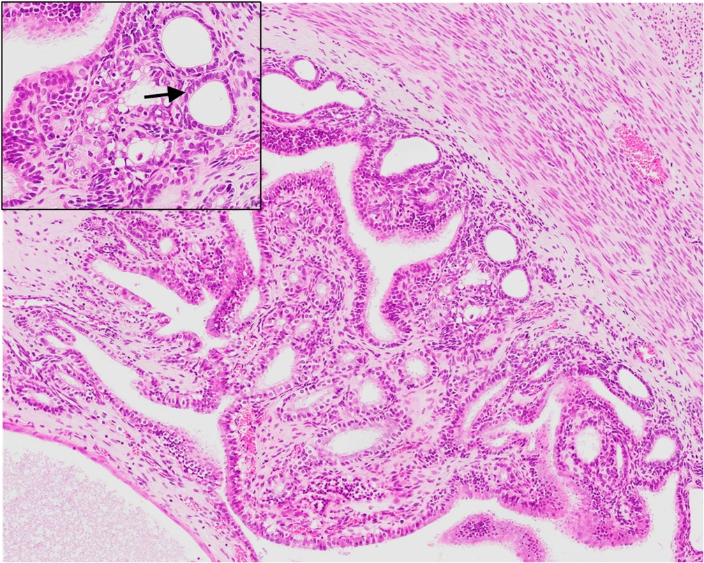 2a
FIGO Grade 1 (Well-Differentiated)
Dominantly glandular or papillary pattern with $\le 5\%$ non-squamous or non-morular solid growth. Minimal nuclear atypia.
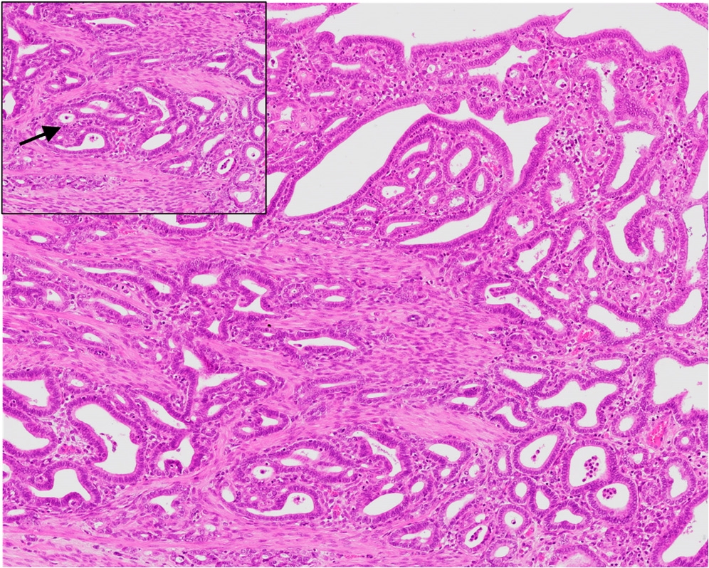 2b
FIGO Grade 2 (Moderately Differentiated)
Intermediate architectural pattern featuring 6% to 50% solid growth pattern mixed with intricate glandular/tubular structures.
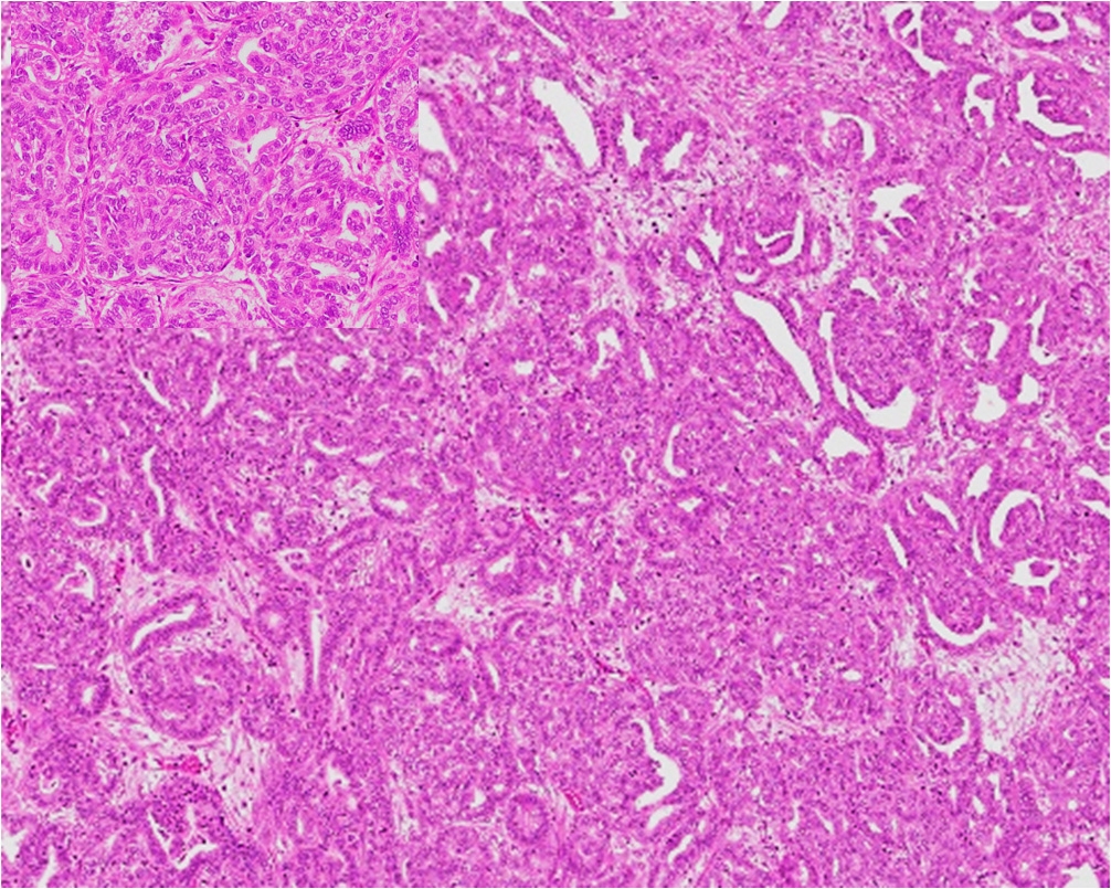 2c
FIGO Grade 3 (Poorly Differentiated)
$>50\%$ solid growth pattern lacking lumen formation, marked cellular atypia, prominent nuclear pleomorphism, and high mitotic index.
"Using human FIGO staging adapted for lagomorph uterine histopathology, tumors were stratified into three tiers: Grade 1 (Figure 2a; 43.3%, 13/30), Grade 2 (Figure 2b; 36.7%, 11/30), and Grade 3 (Figure 2c; 20.0%, 6/30). Grade 1 lesions exhibited low-grade nuclear features with rare mitoses. Grade 2 cases demonstrated progressive loss of glandular architecture and moderate nuclear atypia. Grade 3 tumors were dominated by extensive solid growth ($>50\%$), widespread necrosis, marked nuclear pleomorphism, and brisk mitotic figures ($>5$ per 10 HPF)."
Figure 3 Non-Neoplastic Control Tissues (H&E Staining)
Histological features of healthy rabbit endometrium versus cystic endometrial hyperplasia.
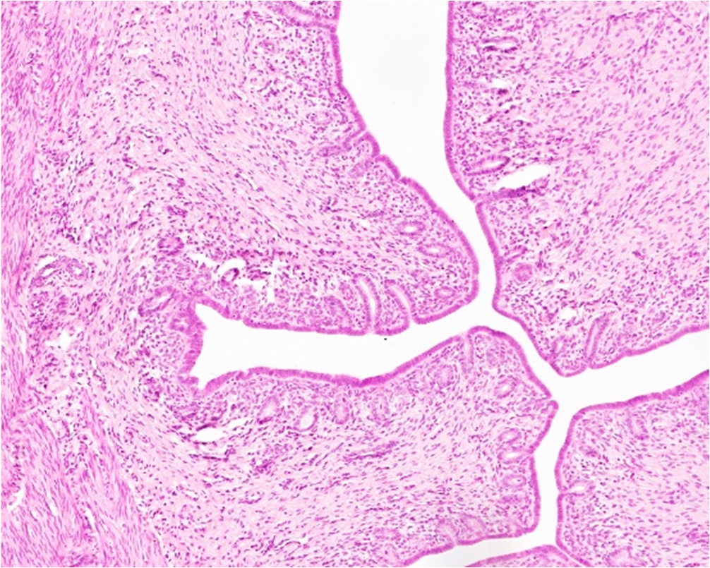 3a
Normal Endometrium
H&E section displaying regular, simple columnar luminal epithelial lining and sparse, round tubular glands within an uninflamed cellular stroma.
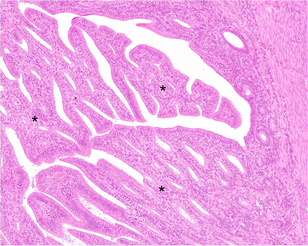 3b
Cystic Endometrial Hyperplasia (CEH)
H&E section showing marked glandular proliferation, back-to-back gland expansion, cystic dilatation of glandular lumens, and hyperplastic columnar epithelium without nuclear atypia.
"Control non-neoplastic tissues comprised normal endometria from young healthy rabbits ($n = 10$; Figure 3a) and cases of cystic endometrial hyperplasia (CEH; $n = 7$; Figure 3b). Normal endometrial tissue presented uniform luminal lining with quiescent tubular glands embedded in compact stroma. In contrast, CEH demonstrated variable degrees of glandular enlargement, papillary infolding, and focal cystic dilatation. Importantly, hyperplastic glands maintained basement membrane integrity without cellular atypia or destructive myometrial invasion."
Figure 4 COX-2 Immunohistochemical Spectrum & Controls
Immunohistochemical staining intensity variations in adenocarcinoma alongside technical controls.
 4a
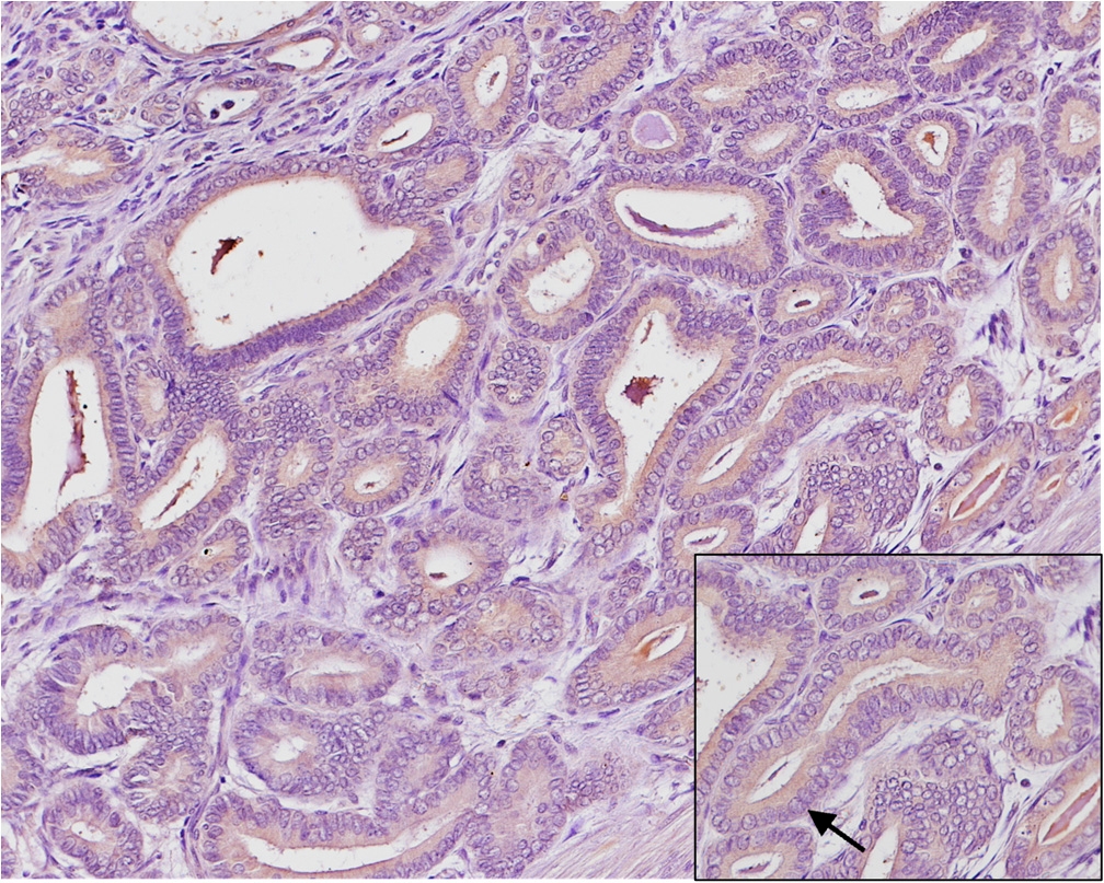
Weak Staining Intensity (Score 1+)
Focal light brown cytoplasm labeling in neoplastic cells (IHS range: 1–4).
 4b
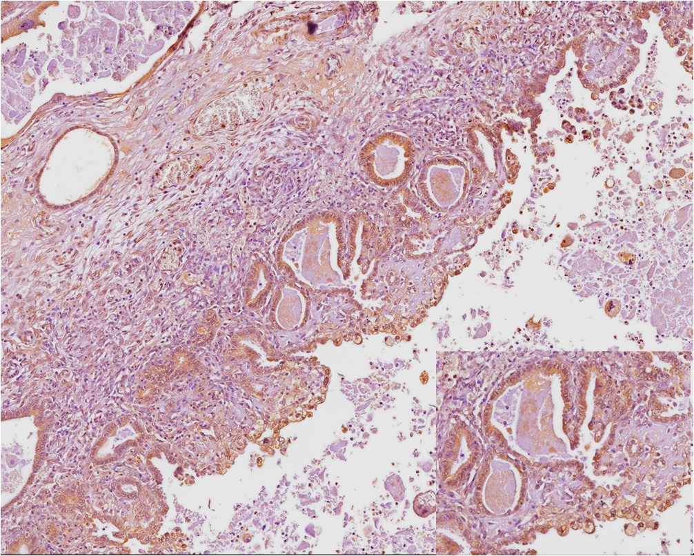
Moderate Staining Intensity (Score 2+)
Distinct, diffuse intermediate cytoplasmic brown staining across tumor glands (IHS range: 6–8).
 4c
Strong / Intense Staining (Score 3+)
Intense granular dark-brown cytoplasmic labeling in virtually all malignant cells (IHS range: 9–12).
 4d
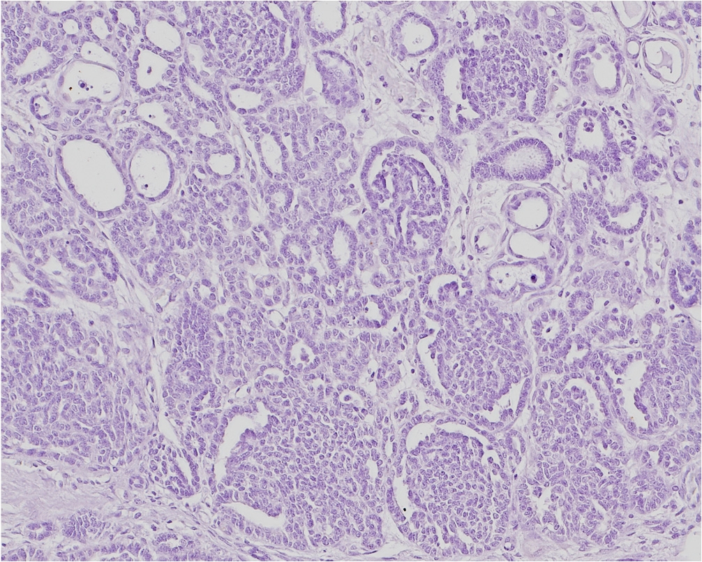
Negative Technical Control
Primary antibody replaced with non-immune isotype mouse IgG, showing zero background chromogen deposition.
 4e
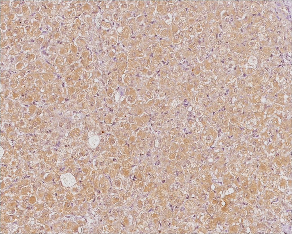
Positive Control (Corpus Luteum)
Strong cytoplasmic COX-2 expression in luteal cells confirming anti-COX-2 antibody cross-reactivity and sensitivity.
"COX-2 expression was quantified using the Immunoreactive Score ($\text{IHS} = \text{Staining Intensity} \times \text{Percentage of Positive Cells}$). Intensity was graded from 0 (negative) to 3 (strong). Percentage of positive cells was scored as 0 (0%), 1 (<10%), 2 (10–50%), 3 (51–80%), and 4 (>80%). All 30 uterine adenocarcinoma cases (100%) were positive for COX-2. Staining intensity ranged from weak (14/30, 46.7%; Figure 4a) to moderate (13/30, 43.3%; Figure 4b) and strong (3/30, 10.0%; Figure 4c). Technical negative controls (Figure 4d) confirmed antibody specificity, while active corpus luteum (Figure 4e) served as positive tissue control."
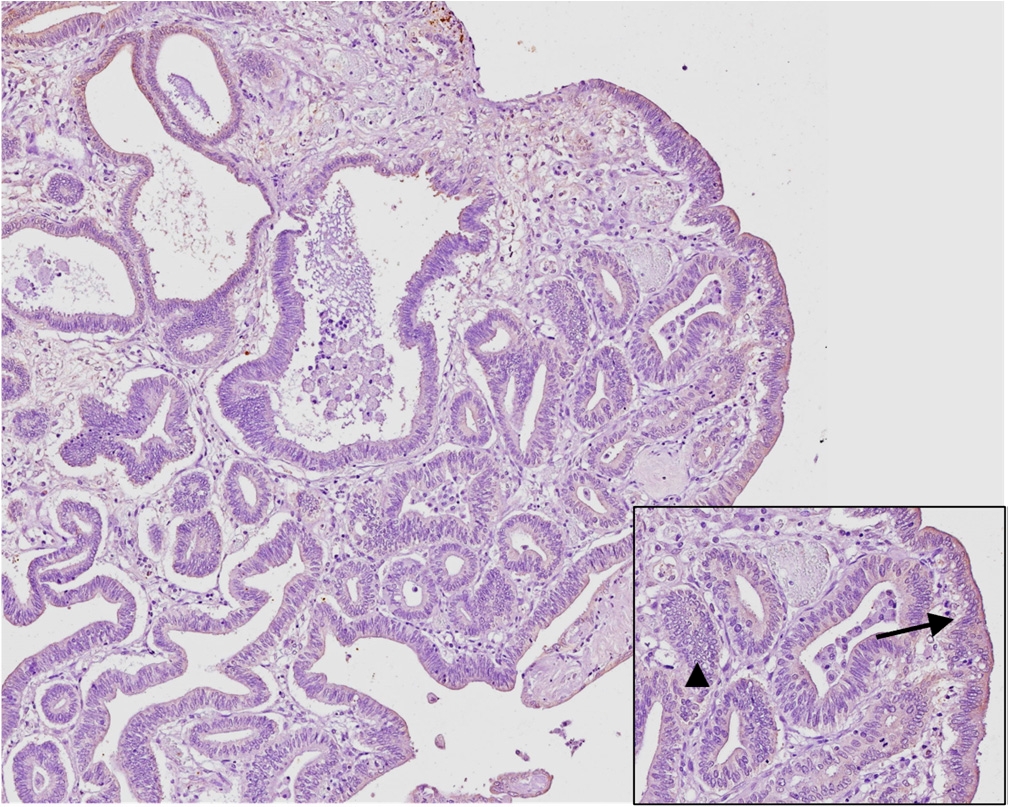
Figure 5 COX-2 Expression in Non-Neoplastic Uterine Controls
Immunohistochemical localization in normal endometrium vs hyperplastic lesions.
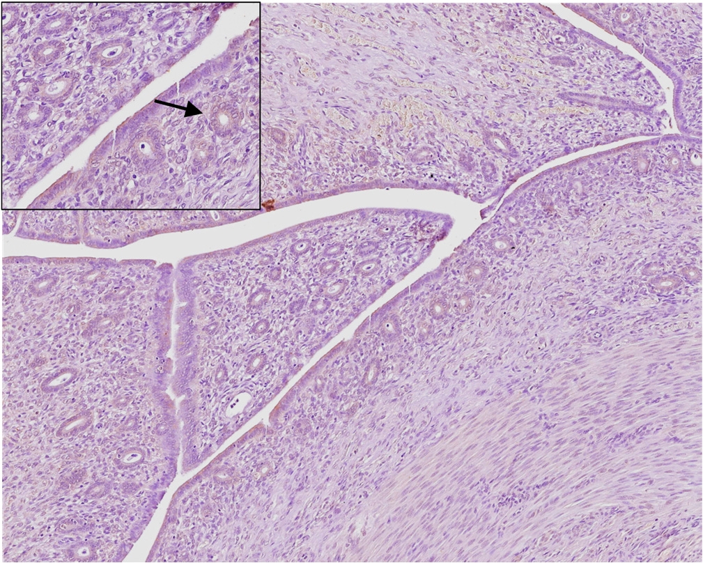 5a
Normal Endometrium (COX-2)
Focal, minimal faint immunostaining strictly limited to the apical border of luminal epithelial cells (arrow), with completely negative stroma and deep glands.
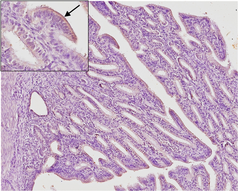 5b
Cystic Endometrial Hyperplasia (COX-2)
Slightly increased low-intensity COX-2 expression localized to the apical cytoplasm of hyperplastic surface epithelium (arrow) and dilated glands.
"In normal endometrial tissue (Figure 5a), COX-2 immunoreactivity was absent or confined to faint apical staining in luminal epithelial cells (mean IHS = $0.40 \pm 0.70$). In cystic endometrial hyperplasia (Figure 5b), weak-to-moderate apical cytoplasmic staining was observed in surface and hyperplastic glandular epithelial cells (mean IHS = $1.71 \pm 1.11$). Stromal cells, myometrium, and vascular endothelium were consistently negative in non-neoplastic specimens."
Figure 6 Quantitative Statistical Evaluation of COX-2 IHS
Statistical analysis comparing COX-2 IHS across tissue categories and histological grades.
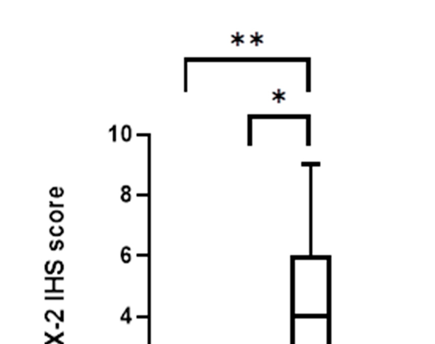
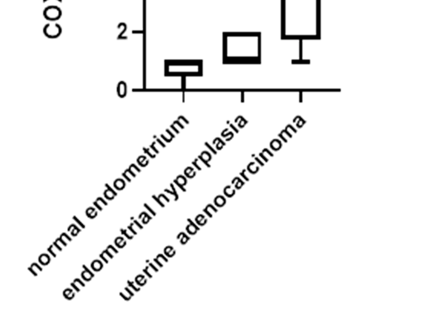
"Statistical analysis revealed a highly significant increase in COX-2 IHS in uterine adenocarcinomas (mean IHS = $8.27 \pm 2.86$) compared to both cystic endometrial hyperplasia (mean IHS = $1.71 \pm 1.11$; $p < 0.001$) and normal endometrium (mean IHS = $0.40 \pm 0.70$; $p < 0.0001$; Figure 6). No significant correlation was observed between COX-2 IHS and FIGO histological grade (Grade 1 mean = 8.15; Grade 2 mean = 8.45; Grade 3 mean = 8.17; $p > 0.05$), indicating that COX-2 overexpression occurs early during malignant transformation and remains consistently high across all evolutionary stages."
Targeted COX-2 Inhibitor Therapy in Rabbit Cancer: Comparative Study of Post-Surgical Meloxicam Treatment vs. Surgery Alone
Detailed comparative clinical follow-up, survival parameters, and molecular mechanisms of Meloxicam action.
Molecular Pathway & Anti-Tumor Mechanism of Meloxicam
Aberrant COX-2 Upregulation in Uterine Epithelium (100% Cases)
Excess PGE₂ Synthesis $\rightarrow$ MMP Activation & Angiogenesis
Meloxicam Inhibition (0.5 mg/kg BID) $\rightarrow$ Blocks PGE₂ Cascade
Apoptosis Induction + Angiogenesis Block $\rightarrow$ >18-Mo Survival
Meloxicam Treatment Protocol ($n = 4$)
A subset of female domestic rabbits ($n = 4$) diagnosed with high-risk uterine adenocarcinoma underwent standard surgical ovariohysterectomy followed by a structured post-operative anti-inflammatory treatment protocol consisting of oral or subcutaneous Meloxicam at 0.5 mg/kg twice daily (BID) for 7 consecutive days.
Histopathological grading confirmed FIGO Grade 2 papillary adenocarcinoma in 3 cases and FIGO Grade 3 tubular/solid adenocarcinoma in 1 case. Despite deep myometrial vascular invasion in 2 cases, 100% of Meloxicam-treated rabbits achieved 18-month metastasis-free survival without evidence of pulmonary recurrence, hematuria, or gastrointestinal toxicity.
Surgery Alone Control Outcomes ($n = 2$)
In contrast, two female rabbits ($n = 2$) presenting with aggressive FIGO Grade 3 tubular/solid uterine adenocarcinoma received ovariohysterectomy alone without post-operative non-steroidal anti-inflammatory drug (NSAID) therapy.
Both control animals experienced rapid disease progression, presenting with dyspnea, weight loss, and multiple pulmonary metastatic nodules confirmed on thoracic radiography. Both rabbits succumbed to metastatic spread at 4 months and 12 months post-surgery respectively (mean survival: 5.4 months).
Clinical Comparison Matrix: Post-Surgical Follow-Up
| Group / Therapy | Sample Size | FIGO Grade | Dosing Regimen | Pulmonary Metastasis | 18-Month Survival | Mean Survival Time |
|---|---|---|---|---|---|---|
| Post-Op Meloxicam Therapy | n = 4 | Grade 2 (3), Grade 3 (1) | 0.5 mg/kg PO BID x 7 days | 0% (0/4) | 100% (4/4) | > 18.0 Months |
| Surgery Alone (No NSAID) | n = 2 | Grade 3 (2) | None (Control) | 100% (2/2) | 0% (0/2) | 5.4 Months |
Full Study Methodology, Results, Discussion, and Reference Index
Abstract
Uterine adenocarcinoma is the most common neoplasm in intact female domestic rabbits (*Oryctolagus cuniculus*), exceeding 60% prevalence in females aged over four years. Cyclooxygenase-2 (COX-2) is an inducible enzyme involved in prostaglandin synthesis, inflammation, and tumor development across multiple species. This study evaluated COX-2 immunoreactivity in 30 cases of rabbit uterine adenocarcinoma, 7 cases of cystic endometrial hyperplasia (CEH), and 10 normal uterine tissue controls. All 30 adenocarcinomas (100%) expressed COX-2 with a mean Immunoreactive Score (IHS) of 8.27 ± 2.86, significantly higher than normal endometrium (0.40 ± 0.70) and CEH (1.71 ± 1.11; p < 0.001). Clinical follow-up demonstrated that post-surgical adjuvant administration of Meloxicam (0.5 mg/kg BID for 7 days) prevented pulmonary recurrence and achieved 100% 18-month survival. These findings establish COX-2 as a critical biomarker and therapeutic target in lagomorph oncology.
Materials and Methods
Sample Selection: Formalin-fixed paraffin-embedded (FFPE) tissue samples from 30 domestic rabbits diagnosed with uterine adenocarcinoma were retrieved from the archives of the Pathology Unit, Department of Veterinary Medical Sciences, University of Bologna. Controls included 10 normal uterine tissues from young healthy rabbits undergoing elective ovariohysterectomy and 7 cases of cystic endometrial hyperplasia (CEH).
Histopathology & FIGO Grading: Sections (3 µm) were stained with Hematoxylin and Eosin (H&E). Tumors were classified into Papillary and Tubular/Solid subtypes and graded according to human FIGO criteria: Grade 1 (≤5% solid growth), Grade 2 (6–50% solid growth), and Grade 3 (>50% solid growth).
Immunohistochemistry (IHC): Antigens were retrieved using citrate buffer (pH 6.0) at 98°C for 20 min. Sections were incubated overnight with a monoclonal mouse anti-COX-2 antibody (clone 44, BD Transduction Laboratories, 1:100 dilution). DAB chromogen was used for visualization. Immunoreactive Score (IHS = Intensity [0–3] × Quantity [0–4]) was calculated (range 0–12).
Results
Histopathology: 16/30 tumors (53.3%) were papillary, and 14/30 (46.7%) were tubular/solid. Superficial myometrial invasion was observed in 18 cases (60%), and deep invasion in 12 cases (40%). FIGO Grade 1 was assigned to 13 cases (43.3%), Grade 2 to 11 cases (36.7%), and Grade 3 to 6 cases (20.0%).
COX-2 Expression: All 30 adenocarcinomas were COX-2 positive. Weak intensity (1+) occurred in 14 cases (46.7%), moderate (2+) in 13 cases (43.3%), and strong (3+) in 3 cases (10.0%). Mean IHS was 8.27 in adenocarcinomas, 1.71 in CEH, and 0.40 in normal endometrium (p < 0.001).
Discussion & Clinical Implications
Overexpression of COX-2 plays a fundamental role in human endometrial cancer progression, facilitating cell survival, invasion, and neoangiogenesis. Our findings confirm that COX-2 upregulation is similarly a hallmark feature of rabbit uterine adenocarcinoma.
The clinical follow-up data demonstrates that post-surgical administration of Meloxicam significantly extends survival and suppresses pulmonary metastasis. Meloxicam provides dual benefits: analgesia/anti-inflammatory control and targeted anti-tumor action by inhibiting PGE₂-driven tumor vascularization.
Full References (Vaccaro et al., 2024)
- Vaccaro, C.; Meoli, S.; Sarli, G.; et al. MDPI Animals 2024, 14, 1318. https://doi.org/10.3390/ani14091318
- Saito, K.; et al. Vet. Pathol. 2002, 39, 299–305.
- Asakawa, M.; et al. J. Vet. Med. Sci. 2008, 70, 811–817.
- Kudo, T.; et al. Cancer Res. 2001, 61, 7307–7314.
- Tschudi, M.; et al. J. Comp. Pathol. 2015, 153, 120–128.